Dr. Jones Research Lab Website

CONTACT US
ANATOMIC ATLAS FOR THE CANINE LUMBOSACRAL SPINE
Dorsal - Mid Canal
Transverse
Sagittal
Dorsal
zoom in
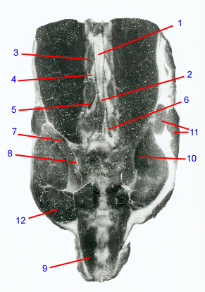
1. Spinal cord.
2. Conus medullaris.
3. Pedicle: paired bony arches that form the lateral boundaries of the vertebral canal. Notches in the caudal and cranial margins of adjacent vertebral pedicles form the cranial and caudal boundaries of the intervertebral foramina, respectively. In dogs that have nerve root compression within the intervertebral foramen, portions of the caudal pedicle often have to be removed.
4. Intervertebral foramen: paired, "horse-head" shaped openings between adjacent vertebrae that form the passageway through which the nerve roots exit the vertebral canal. Boundaries consist of the articular processes dorsally, pedicles cranially and caudally, and the vertebral endplates/disc ventrally. In dogs with degenerative lumbosacral disease, nerve roots and their associated blood vessels can become compressed or entrapped within the intervertebral foramina.
5. L6 nerve roots lead to the femoral, obturator, cranial gluteal, and sciatic peripheral nerves-- innervating the iliopsoas, quadriceps, and sartorius muscles, the external obturator, pectineus, gracilis, and adductor muscles, the middle gluteal, deep gluteal, and tensor fascia lata muscles, and the biceps femoris, semimembranosus muscles.
6. L7 nerve roots lead to the cranial gluteal, caudal gluteal, and sciatic peripheral nerves. The cranial and caudal gluteal peripheral nerves innervate the middle gluteal, deep gluteal, and tensor fascia lata muscles, and the superficial gluteal muscles. The sciatic peripheral nerve innervates the biceps femoris, semimembranosus, and semitendinosus muscles, as well as the common peroneal nerve (which innervates the peroneus longus, lateral digital extensor, long digital extensor, and cranial tibial muscles), and the tibial nerve (which innervates the gastrocnemius, popliteus, superficial digital flexor, and deep digital flexor muscles).
7. Wing of ilium.
8. Sacroiliac joint: partly cartilaginous and partly synovial. Permits rotational and lateral movement of the pelvis relative to the sacrum. Can develop osteoarthritis and become a source of pain in the lower back region. Dogs with congenital lumbosacral vertebral anomalies can also have assymmetrical sacroiliac joints.
9. Coccygeus.
10. Gluteus medius.
11. Sartorius.
12. Gluteus superficialis.
Go to top