Dr. Jones Research Lab Website

CONTACT US
ANATOMIC ATLAS FOR THE CANINE LUMBOSACRAL SPINE
Dorsal - Lamina
Transverse
Sagittal
Dorsal
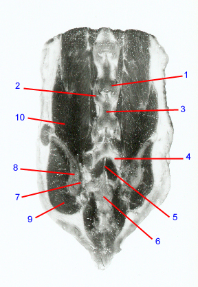
1. Articular process: Synonym: facet. Interdigitating bony prominences that form the articular process joints between adjacent vertebrae. In the lumbar spine, these form the dorsal boundaries of the intervertebral foramina. In dogs that have nerve root compression within the intervertebral foramen, portions of the articular processes often have to be removed.
2. L6 cranial articular process
3. Lamina: forms the dorsal boundary of the vertebral canal. Has small notches on the cranial and caudal margins that form the boundaries of the interlaminar spaces and serve as the attachment sites for the yellow (interarcuate) ligaments. In dogs with idiopathic stenosis, the laminae may appear thickened and sclerotic .
4. L7 caudal articular process
5. Interlaminar space: Synonym: interarcuate space. Space between laminae of adjacent vertebrae. Occupied by the yellow (interarcuate) ligament and medial portions of the articular process joint capsules. In dogs with degenerative lumbosacral disease, the L7-S1 interlaminar space is often narrowed. This is associated with buckling and hypertrophy of the ligaments.
6. Body of sacrum: formed by the fusion of the S1-3 vertebral bodies. Ventral subluxation of the sacrum can contribute to compression of nerve roots in dogs with degenerative lumbosacral disease.
7. Sacroiliac joint: partly cartilaginous and partly synovial. Permits rotational and lateral movement of the pelvis relative to the sacrum. Can develop osteoarthritis and become a source of pain in the lower back region. Dogs with congenital lumbosacral vertebral anomalies can also have assymmetrical sacroiliac joints.
8. Wing of ilium
9. Erector spinae
10. Gluteus medius
Go to top