Dr. Jones Research Lab Website

CONTACT US
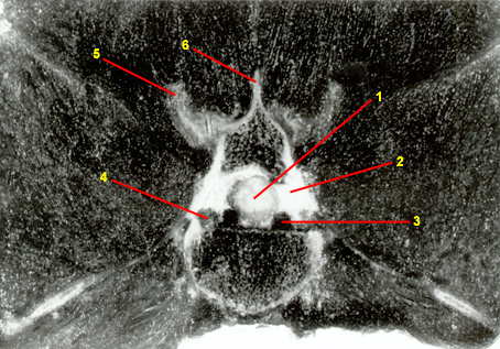
ANATOMIC ATLAS FOR THE CANINE LUMBOSACRAL SPINE
Transverse - Cranial L6
Transverse
Sagittal
Dorsal
(Jones et al., 1995)
1. Thecal sac: Synonym dural sac. Soft tissue opacity CT structure that is enclosed by the dura mater. Tissues inside the thecal sac include the arachnoid membrane, fluid-filled subarachnoid space, nerve roots, and spinal cord. Individual soft tissues inside of the thecal sac are silhouetted in plain CT images and cannot be discriminated.
2. Epidural fat: fat deposits are normally present in the epidural space and appear darker than surrounding nerve tissues on CT images. These fat deposits help provide cushioning and protection of the nerve tissues. In dogs with degenerative lumbosacral disease, loss of visualization of the epidural fat is a sign of nerve tissue compression.
3. Internal vertebral venous plexus.
4. L5 nerve roots lead to the femoral and obturator peripheral nerves-- innervating the iliopsoas, quadriceps, and sartorius muscles, and the external obturator, pectineus, gracilis, and adductor muscles.
Nerve root: paired bundles of nerve tissue that exit the spinal cord at each spinal cord segment. Surrounded by dura mater, arachnoid membrane and cerebrospinal fluid. Consists of ventral and dorsal spinal nerves, and a dorsal root ganglion.
5. Mammillary process: Synonym metapophysis. Small, knoblike bony eminence visible on the dorsal margins of the transverse processes in the cranial thoracic region and the cranial articular processes in the caudal thoracic-coccygeal regions. Can sometimes be misinterpreted as osteophytes in dogs with suspected spinal osteoarthritis.
6. Spinous process: dorsally projecting bony prominence arising from the center of each vertebral lamina. Serves as an attachment site for epaxial muscles and the interspinous ligament. The lamina of L7 and the L7 spinous process are often removed for lumbosacral decompressive surgeries.
Go to top