Dr. Jones Research Lab Website

CONTACT US
ANATOMIC ATLAS FOR THE CANINE LUMBOSACRAL SPINE
Transverse
Sagittal
Dorsal
Sagittal - Central Canal Discs
zoom in
1. Multifidus muscles: one of the largest epaxial muscle groups in the lumbar region. Divided into 11 individual portions that orginiate from the articular processes of the sacrum and mammillary processes of T12-L7. Can appear atrophied in dogs with chronic degenerative spinal disease or degenerative myelopathy.
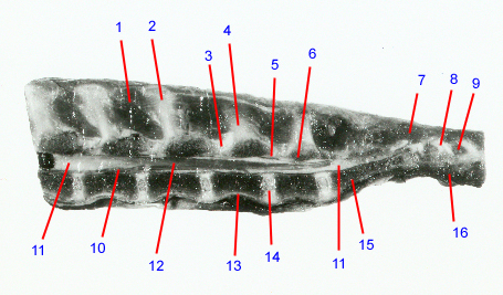
2. Spinous process: dorsally projecting bony prominence arising from the center of each vertebral lamina. Serves as an attachment site for epaxial muscles and the interspinous ligament. The lamina of L7 and the L7 spinous process are often removed for lumbosacral decompressive surgeries.
3. Interlaminar space: Synonym: interarcuate space. Space between laminae of adjacent vertebrae. Occupied by the yellow (interarcuate) ligament and medial portions of the articular process joint capsules. In dogs with degenerative lumbosacral disease, the L7-S1 interlaminar space is often narrowed. This is associated with buckling and hypertrophy of the ligaments.
4. L6 spinous process
5. Yellow ligament: Synonyms ligamentum flavum, interarcuate ligament. Loose, thin, elastic sheets between the laminae of adjacent vertebrae. They blend into the joint capsules of the articular processes. In dogs with degenerative lumbosacral stenosis, the yellow ligament at L7-S1 can become thickened or hypertrophied. When this happens, it can cause pain and weakness due to compression of the cauda equina nerve roots.
6. Lamina: forms the dorsal boundary of the vertebral canal. Has small notches on the cranial and caudal margins that form the boundaries of the interlaminar spaces and serve as the attachment sites for the yellow (interarcuate) ligaments. In dogs with idiopathic stenosis, the laminae may appear thickened and sclerotic.
7. Sacrocaudalis dorsalis medialis
8. Pedicle: paired bony arches that form the lateral boundaries of the vertebral canal. Notches in the caudal and cranial margins of adjacent vertebral pedicles form the cranial and caudal boundaries of the intervertebral foramina, respectively. In dogs that have nerve root compression within the intervertebral foramen, portions of the caudal pedicle often have to be removed.
9. Intervertebral foramen: paired, “horse-head” shaped openings between adjacent vertebrae that form the passageway through which the nerve roots exit the vertebral canal. Boundaries consist of the articular processes dorsally, pedicles cranially and caudally, and the vertebral endplates/disc ventrally. In dogs with degenerative lumbosacral disease, nerve roots and their associated blood vessels can become compressed or entrapped within the intervertebral foramina.
10. Internal vertebral venous plexus
11. Epidural fat: fat deposits are normally present in the epidural space and appear darker than surrounding nerve tissues on CT images. These fat deposits help provide cushioning and protection of the nerve tissues. In dogs with degenerative lumbosacral disease, loss of visualization of the epidural fat is a sign of nerve tissue compression.
12. Spinal cord
13. Ventral longitudinal ligament
14. Intervertebral disc: Cartilaginous joint between endplates of adjacent vertebrae. Consists of concentric bands of fibrous tissue (annulus fibrosus) surrounding a central gelatinous portion (nucleus pulposus). Type II disc degeneration is one of the most common causes of nerve root compression in dogs with degenerative lumbosacral disease. With type II disc degeneration, the annulus bulges outwardly in all directions and bone spurs form on the vertebral endplates at the attachment sites for the annulus.
15. Body of sacrum: formed by the fusion of the S1-3 vertebral bodies. Ventral subluxation of the sacrum can contribute to compression of nerve roots in dogs with degenerative lumbosacral disease.
16. Sacrocaudalis ventralis lateralis
Go to top