Dr. Jones Research Lab Website

CONTACT US
ANATOMIC ATLAS FOR THE CANINE LUMBOSACRAL SPINE
Dorsal - Articular Process Joints
Transverse
Sagittal
Dorsal
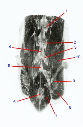
1. Articular process joint: Paired, synovial joints dorsal to the vertebral canal. Formed by the cranial and caudal articular processes of contiguous vertebrae. Osteoarthritis of the articular process joints can contribute to nerve root compression in dogs with degenerative lumbosacral disease.
2. Articular process: Synonym: facet. Interdigitating bony prominences that form the articular process joints between adjacent vertebrae. In the lumbar spine, these form the dorsal boundaries of the intervertebral foramina. In dogs that have nerve root compression within the intervertebral foramen, portions of the articular processes often have to be removed.
3. L6 caudal articular process
4. Lamina: forms the dorsal boundary of the vertebral canal. Has small notches on the cranial and caudal margins that form the boundaries of the interlaminar spaces and serve as the attachment sites for the yellow (interarcuate) ligaments. In dogs with idiopathic stenosis, the laminae may appear thickened and sclerotic.
5. Spinous process: dorsally projecting bony prominence arising from the center of each vertebral lamina. Serves as an attachment site for epaxial muscles and the interspinous ligament. The lamina of L7 and the L7 spinous process are often removed for lumbosacral decompressive surgeries.
6. Body of sacrum: formed by the fusion of the S1-3 vertebral bodies. Ventral subluxation of the sacrum can contribute to compression of nerve roots in dogs with degenerative lumbosacral disease.
7. Multifidus muscles: one of the largest epaxial muscle groups in the lumbar region. Divided into 11 individual portions that orginiate from the articular processes of the sacrum and mammillary processes of T12-L7. Can appear atrophied in dogs with chronic degenerative spinal disease or degenerative myelopathy.
8. Gluteus medius
9. Wing of ilium
10. Erector spinae
Go to top