Dr. Jones Research Lab Website

CONTACT US
ANATOMIC ATLAS FOR THE CANINE LUMBOSACRAL SPINE
Transverse - L7-S1 Disc - Zoom
Transverse
Sagittal
Dorsal
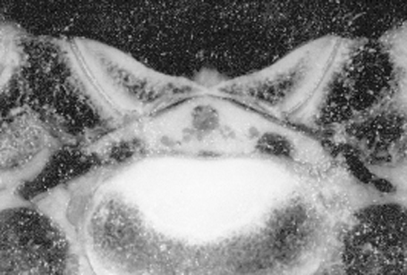
(Jones et al., 1995)
zoom out
1. Thecal sac: Synonym dural sac. Soft tissue opacity CT structure that is enclosed by the dura mater. Tissues inside the thecal sac include the arachnoid membrane, fluid-filled subarachnoid space, nerve roots, and spinal cord. Individual soft tissues inside of the thecal sac are silhouetted in plain CT images and cannot be discriminated.
2. Epidural fat: fat deposits are normally present in the epidural space and appear darker than surrounding nerve tissues on CT images. These fat deposits help provide cushioning and protection of the nerve tissues. In dogs with degenerative lumbosacral disease, loss of visualization of the epidural fat is a sign of nerve tissue compression.
3. Internal vertebral venous plexus
4. S1 nerve roots: lead to the cranial gluteal, caudal gluteal, sciatic, and pudendal peripheral nerves. The cranial gluteal peripheral nerve innervates the middle gluteal, deep gluteal, and tensor fascia lata muscles. The caudal gluteal peripheral nerve innervates the superficial gluteal, and middle gluteal muscles.
5. S2 nerve roots lead to the caudal gluteal, sciatic, and pudendal peripheral nerves. The caudal gluteal peripheral nerve innervates the superficial gluteal, and middle gluteal muscles.
6. S3 nerve root leads to the pudendal peripheral nerve. The pudendal peripheral nerve innervates the the caudal rectal nerve (which innervates the external anal sphincter muscle).
7. Yellow ligament: Synonyms ligamentum flavum, interarcuate ligament. Loose, thin, elastic sheets between the laminae of adjacent vertebrae. They blend into the joint capsules of the articular processes. In dogs with degenerative lumbosacral stenosis, the yellow ligament at L7-S1 can become thickened or hypertrophied. When this happens, it can cause pain and weakness due to compression of the cauda equina nerve roots.
8. Spinal nerve: Paired, segmental nerves that connect the spinal cord to peripheral structures. In the dog, there are usually 36 pairs. Each spinal nerve consists of 4 components: 1) roots, 2) main trunk, 3) four primary branches, and 4) numerous peripheral branches.
9. Intervertebral vein: branch of the ventral vertebral venous plexus that exits the caudal portion of the intervertebral foramen and empties into the caudal vena cava. Can become congested in dogs with degenerative disc disease and contribute to compression of nerve roots in the intervertebral foramen. Believed to be an important factor in a syndrome called “intermittent claudication”, where lameness arises during exercise and resolves after rest.
10. Sacroiliac joint: partly cartilaginous and partly synovial. Permits rotational and lateral movement of the pelvis relative to the sacrum. Can develop osteoarthritis and become a source of pain in the lower back region. Dogs with congenital lumbosacral vertebral anomalies can also have assymmetrical sacroiliac joints.
11. Wing of ilium
12. Wing of sacrum: elongated lateral process off S1 that articulates with the ilium to form the sacroiliac joint. The sacrum is formed by the fusion of the S1-3 vertebrae. It has paired dorsal and ventral foramina at S1-2 and S2-3 junctions where the dorsal and ventral branches of S1 and S2 nerve roots exit.
13. Multifidus muscles: one of the largest epaxial muscle groups in the lumbar region. Divided into 11 individual portions that orginiate from the articular processes of the sacrum and mammillary processes of T12-L7. Can appear atrophied in dogs with chronic degenerative spinal disease or degenerative myelopathy.
Go to top