Dr. Jones Research Lab Website

CONTACT US
ANATOMIC ATLAS FOR THE CANINE LUMBOSACRAL SPINE
Sagittal - L Sacroiliac Joint
Transverse
Sagittal
Dorsal
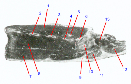
1. Sacrocaudalis dorsalis lateralis
2. Transverse process: paired bony prominences that project laterally from the region where the pedicle joins the vertebral body. In the lumbar region, they project cranially and ventrally. Serve as attachment sites for paraspinal muscles. Ventral branches of the spinal nerves course ventral to these processes.
3. L6 transverse process
4. L7 transverse process
5. Articular process: Synonym: facet. Interdigitating bony prominences that form the articular process joints between adjacent vertebrae. In the lumbar spine, these form the dorsal boundaries of the intervertebral foramina. In dogs that have nerve root compression within the intervertebral foramen, portions of the articular processes often have to be removed.
6. Wing of sacrum: elongated lateral process off S1 that articulates with the ilium to form the sacroiliac joint. The sacrum is formed by the fusion of the S1-3 vertebrae. It has paired dorsal and ventral foramina at S1-2 and S2-3 junctions where the dorsal and ventral branches of S1 and S2 nerve roots exit.
7. Psoas major
8. Quadratus lumborum
9. Retroperitoneal fat
10. Sacroiliac joint: partly cartilaginous and partly synovial. Permits rotational and lateral movement of the pelvis relative to the sacrum. Can develop osteoarthritis and become a source of pain in the lower back region. Dogs with congenital lumbosacral vertebral anomalies can also have assymmetrical sacroiliac joints.
11. Iliopsoas
12. Quadratus femoris
13. Quadratus lumborum
Go to top